Echocardiogram
AN ULTRASOUND TO CREATE PICTURES OF THE HEART
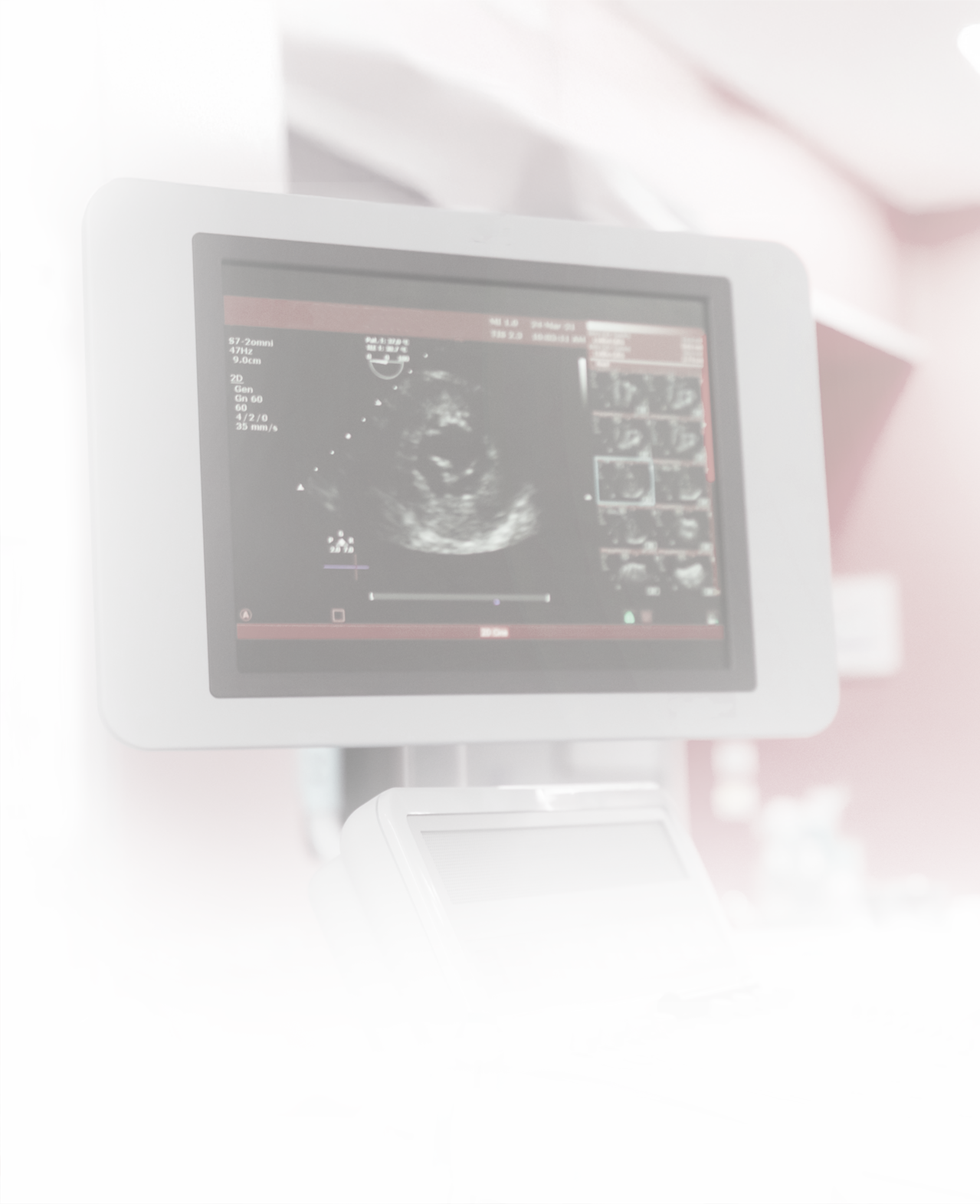
An echocardiogram uses sound waves to produce images of the heart. This test allows your doctor to see how well your heart is squeezing and to identify abnormalities with the heart muscle and valves.
There is no special preparation for this test. You will be asked to undress from the waist up. You will then be covered with a gown and asked to lie slightly on your left side. EKG electrodes will be connected to your chest. Using a small amount of ultrasound gel and a transducer placed over and around your heart, your doctor will take images of your heart. You may be asked to take deep breaths, and sometimes the transducer must be held firmly against your chest. This may cause a little discomfort but produces the best pictures of your heart.
THIS PROCEDURE TAKES 30-45 MINUTES. WHEN YOU ARRIVE FOR THIS PROCEDURE, PLEASE BRING THE FOLLOWING ITEMS WITH YOU:
All your current medications in their original containers
Your current picture ID
Your current insurance card

